 |
| Photo Credit: youtube |
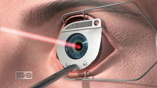
The use of laser
technology has increased greatly years and lasers have many applications in
engineering, science, medicine and the defense industry. A laser beam is a
monochromatic, coherent, parallel, very high energy beam.
It is produced by the
input of radiant energy, which causes the emission of light as the atoms return
to their original energy state. Many substances can be used as a laser source,
e.g. ruby, argon, carbon dioxide, or YAG (yttrium-aluminium-garnet). 'Laser'
is an acronym for 'Light Amplification by Stimulated Emission of Radiation'.
Lasers have been produced to emit at many monochromatic wavelengths ranging
from far UV (less than 300nm) to IR (greater than 1400nm).
Continuous wave laser
systems, such as argon and krypton lasers, focus light energy on an area of
tissue to cause a temperature rise, which in turn causes coagulation of the
tissue. The temperature rise depends on the spot size, amount of energy and
wavelength. The wavelength determines the efficiency with which the light
source is absorbed and converted into heat by the pigment of the tissues.
An excessive temperature
rise can cause an explosion and hemorrhage in the tissue. Only a 10C rise in
temperature is required for retinal photocoagulation. Short pulse lasers, such
as YAG, will disrupt transparent tissue and hence are known as photodisruptors.
They are generally used for the relatively transparent anterior portion of the
eye.
There are various
mechanisms whereby the short pulse laser systems disrupt the transparent
membranes:
- The high irradiance (power/area) disintegrates tissues by removing electrons from atoms (i.e. ionizing them) and creating a 'plasma', which is a gaseous state consisting of electrons and ions.
- The rapid outwards expansion of the plasma creates shock and acoustic waves, which mechanically disrupt adjacent tissue.
- Latent stress in the membrane causes further disruption.
- A specific photochemical reaction has been reported with UV lasers, which results in the ablation of corneal tissues without thermal damage to the adjacent structures.
The site of absorption
depends upon the wavelength being emitted by the laser. Radiation from lasers
emitting in the UV region will be absorbed by the cornea and crystalline lens.
Vision and IR lasers will cause problems, not necessarily because of their
power but due to the fact that the collimated beam will be focused on the
retina.
UV lasers emitting
radiation less than 300nm can be absorbed by the cornea and, as described
above, will cause damage to the corneal epithelium. Lasers emitting UV from
300-400nm can cause cataracts because the radiation is absorbed by the
crystalline lens. UV-emitting lasers can therefore cause damage to the cornea
and crystalline lens, if they are exposed for a sufficient time and at power
densities above threshold level. As a result of this, the Excimer laser is now
being used, particularly for corneal surgery, as the depth of incision can be
accurately controlled by the photons that break the molecular bonds when
absorbed. This has the advantage that the adjacent tissue does not suffer from
thermal damage.
It's has been observed
that the wavelength of 193nm is optimal for corneal surgery. Excimer laser
was initially used in the printing and electronics industries, where its
properties allowed submicron patterns to be etched into the surface of
materials without damaging the adjacent non-irradiated areas.
The wavelengths of
visible and IR lasers are focused on to the retina, where they cause damage:
- Thermal injury; Absorption by the melanin, the pigment epithelium and choroid.
- Photochemical damage; Especially in the blue region of the visible spectrum, which is absorbed by the inner retinal layers.
- Shock waves; These are produced by the Q-switched high intensity beams, which cause disruption of the internal cellular structure.
The type and degree of
retinal damage caused by a laser will depend upon the following:
- The power density of the laser;
- Time of exposure;
- The wavelength and transmission through the ocular
media;
- The size of image upon the retina;
- The blink reflex;
- The degree of retinal and choroidal pigmentation.
Industrial ocular laser burns have been reported. "The damage ranged from minor retinal burns to extensive areas of damage with retinal oedema and vitreous hemorrhages. A variety of lasers were involved, including YAG, argon, krypton and rhodamine dye. The hemorrhage and oedema subsided during the weeks following the injury and with the exception of one case; the final visual acuity was 6/6, although a paracentral scotoma was usually present.
In all but one case eye
protection was not being worn; in the case where eye protection was being worn,
it was not fitted correctly and the spectacles slipped as the worker bent over
the laser, the beam being reflected from a piece of test paper into his left
eye. There are other reports of ocular injury from lasers where the visual
outcome was not as good. Many of the cases resulted in a markedly reduced final
vision, one case being due to a macula hole" (Jacobson et al.)

No comments:
Post a Comment
We love to hear from you!
Please share your thought here!