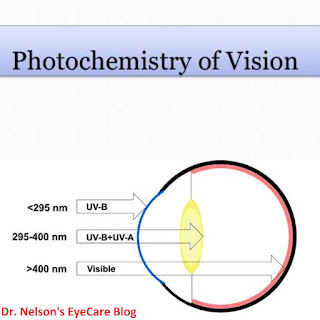
Before light reaches the retina it must pass through the optical media of the eye (cornea, aqueous humor, lens and vitreous humor).
When this light impinges the retina, the photoreceptors are excited and this is where the photochemistry of vision begins. Photochemistry of vision does not only tell us how vision is perceived rather it also exposes the photochemical and biochemical changes occurring in the retina and the visual transduction of the impulses generated from these changes to the brain for interpretation.
When this light impinges the retina, the photoreceptors are excited and this is where the photochemistry of vision begins. Photochemistry of vision does not only tell us how vision is perceived rather it also exposes the photochemical and biochemical changes occurring in the retina and the visual transduction of the impulses generated from these changes to the brain for interpretation.
`
Photochemical Change
When visible light hits the rhodopsin, the 11-cis-retinal (which fits perfectly in the protein component of rhodopsin) undergoes an isomerization in molecular arrangement to form all-trans-retinal which does not fit well in the protein. The inability of all-trans-retinal to fit well in the protein leads to a series of conformational changes in the protein. As the protein changes its conformation, it initiates a cascade of biochemical reaction that result in the closing of sodium ion channels in the cell membrane.
Before the closure, sodium ions flow freely into the cell to compensate for the lower potential (more minus charge) which exists inside the cell. This potential difference is passed along an adjoining nerve cell as an electrical impulse at the synaptic terminal. The nerve cell carries this impulse to the brain, where the visual information is interpreted.
Biochemical Change
When rhodopsin absorbs a photon it isomerizes to all-trans configuration without any accompanying change in the structure of the protein. Rhodopsin containing the all trans isomer of retinal is known as bathorhopsin. However, the trans isomer does not fit well into the protein due to its rigid elongated shape. Therefore a series of changes occurs to expel the all-trans-retinal from the protein.
The configuration of all-trans-retinal in bathorhodopsin is too unstable to remain in this configuration for long and within a nanoseconds, the shape of the protein begins to change. The bathorhodopsin undergoes a conformational change to produce a series of intermediate complexes (Lumirhodopsin, metarhodopsin1 and metarhodopsin2) each absorbing light maximally at a different wavelength. The important intermediate metarhodopsin2 releases the retinal as free trans-retinal and then activates the enzyme tranducin which initiates the visual transduction cascade.
The tranducin enzyme activates another enzyme called phosphodiesterase which hydrolyzes cyclic GMP. Scarcity of cyclic GMP closes the sodium ion channels and this closure builds up a large potential difference across the plasma membranes. The potential difference is transmitted to the brain as an electrical impulse. The free trans-retinal is regenerated to 11-cis-retinal and re-incorporated into rhodopsin in dim light when the photon is removed.


No comments:
Post a Comment
We love to hear from you!
Please share your thought here!